Hemofilia typu A i B u psów
Clinical signs of canine haemophilia A and B
The haemophilic phenotype has been observed in many dog breeds and in mixed breed dogs (Brooks, 1999; Mischke, 2012). Because these haemostasis disorders are transmitted as an X-linked trait, male dogs are clinically affected while the bitches normally act as asymptomatic carriers (Benn et al., 1978; Johnstone et al., 1984; Mischke, et al., 2011C; Mischke, 2012).
The clinical manifestation of haemophilia A and B is very similar (Peterson et al., 1979; Mischke, 2012). Clinical signs usually appear at an early age (Campbell et al., 1983). Commonly, repeated episodes of bleeding occur at birth from the umbilical cord (Gentry et al., 1977; Benn et al., 1978; Johnstone el al., 1984) and during teething (Gentry et al., 1977; Benn et al., 1978; Peterson et al., 1979; Campbell et al., 1983; Brooks et al., 1997; Iazbik et al., 1997; Nakata et al., 2006).
Affected dogs may also show prolonged bleeding from cutaneous wounds (Brooks et al., 2003; Mischke et al., 2011C) and excessive bleeding after trauma (Gentry et al., 1977; Benn et al., 1978; Brooks et al., 2003), minimal surgical procedures or medical interventions (Gentry et al., 1977; Verlander et al., 1984; Dunning et al., 2009; Mischke et al., 2011C). Spontaneous musculoskeletal bleeding has been also described in severely affected dogs (Gentry et al., 1977; GU et al., 1999; Hough et al., 2002; Mischke et al., 2011C). In addition, subcutaneous (Sugahara et al., 1996; Brooks et al., 1997; Nakata et al., 2006) or intramuscular haematoma formations are also frequent (Peterson et al., 1979; Feldman et al., 1995; Nakata et al., 2006). Depending on the position and extension of the haemorrhages and haematomas, affected dogs may show different signs.
The severity of haemophilia varies depending on the type of mutation in the FVIII/FIX gene (Feldman et al., 1995), on the in vitro measurement of the residual FVIII (Johnstone et al., 1984) or FIX coagulant activity (Feldman et al., 1995), and on clinical factors including the size of the breed, the dog activity (Mischke, 2012), and keeping conditions (Nakata et al., 2006). Some authors classify the severity of haemophilia based on human criteria (Benn et al., 1978; Johnstone et al., 1984; Nakata et al., 2006). However it is known that the haemophilic dogs show more severe signs than haemophilic human at the same reduced FVIII/FIX activity. These differences in the expression of haemophilia between both species have been related to different factors, such as the size of the breed and the dog activity (Mischke, 2012).
Diagnosis of canine haemophilia A and B
Haemophilia A and B should be suspected in bitches with a family history of bleeding disorder and/or in male dogs with a bleeding history (Feldman et al., 1995).
Affected male dogs show normal buccal mucosal bleeding time, prolonged cuticle bleeding time with re-bleeding (Sherding et al., 1980; Brooks et al., 1997; Nakata et al., 2006), as well as prolonged activated partial thromboplastin time (APTT) and activated clotting time, because reduced factor VIII/FIX activity impairs clot formation via the intrinsic pathway of coagulation (Feldman et al., 1995). It should be noted that APTT can be normal due to a stress response to the bleeding crisis, which increases the fibrinogen concentration and shortens the time of clot formation (Campbell et al., 1983, Mischke, 2000).
To confirm the diagnosis, specific measurements of factor activity have to be performed (Brooks et al., 2003; Mischke, 2012). Affected animal normally show an activity of the haemophilia factor less than 10%, but in mild forms it can be 10-25% (Mischke, 2012). In haemophilic dogs, factor concentration can also be measured (Brooks et al., 1997; Gu et al., 1999; Hough et al., 2002). However, in case of a structural change of the protein, the immunologic assay may lead to wrong results, due to the possible alteration of antibody binding sites on the protein (Mischke, 2012).
The diagnosis of asymptomatic carrier should be based on the pedigree analysis and specific genetic tests (Mischke, 2012). Due to the additional influence of other clotting factors, the APTT assay is not reliable for the detection of carriers due to (Johnstone et al., 1984; Mischke, 2012). In addition, although carriers normally have about 50% of normal FVIII activity (Benn et al., 1978; Johnstone et al., 1984; Mischke, 2012) or less than 50% (Sugahara et al., 1996; Mischke, 2012), there is an overlap between carrier and healthy dogs, which complicates the diagnose (Benn et al., 1978; Johnstone et al., 1984).
The calculation of the vWF concentration/FVIII activity ratio, which is usually elevated (>2.00) in carrier dogs, can help to find the diagnosis (Johnstone et al., 1984; Mischke, 2012). Currently, the identification of mutations in some dog breeds has enabled the elaboration of genetic tests (Tab. 1). These genetic tests allow us to diagnose female carriers of the trait, which is crucial for the control of haemophilia (Mischke et al., 2011A; Mischke et al., 2011B; Mischke et al., 2011C).
Mogą zainteresować Cię również


Znajdź swoją kategorię
2609 praktycznych artykułów - 324 ekspertów - 22 kategorii tematycznych


Weterynaria w Terenie
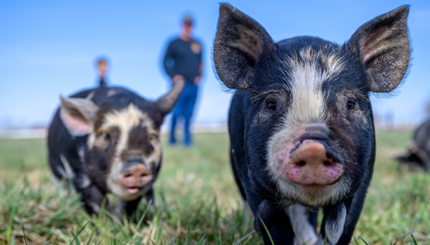
Praktyczne aspekty diagnostyki laboratoryjnej chorób świń
Strategia DIVA Kolejnym problemem jest różnicowanie serologiczne w przypadku wyniku pozytywnego, czy świnie były szczepione przeciw określonym chorobom lub czy są bezobjawowymi nosicielami chorobotwórczego wirusa. W tym celu opracowane zostały specjalne testy, które umożliwiają odróżnienie przeciwciał wywołanych przez wirus szczepionkowy specjalnie w tym celu genetycznie modyfikowany od przeciwciał wytworzonych pod wpływem wirusa zjadliwego. Powyższe określane […]


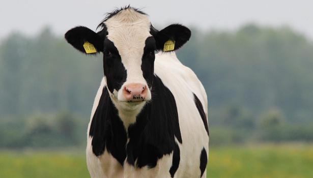
Podkliniczny niedobór wapnia i jego wpływ na wyniki rutynowych badań laboratoryjnych w różnych okresach laktacji u krów mlecznych
Diagnostyka serologiczna Badania serologiczne są najwygodniejszym i najtańszym sposobem identyfikacji stad bezobjawowo zakażonych App. W badaniach tych rutynowo wykorzystuje się testy ELISA. W odniesieniu do App rozróżnia się 2 typy tych testów. Pierwszy (ze względu na cenę najczęściej wykorzystywany) bazuje na wytwarzanej przez wszystkie 15 serotypów toksynie Apx IV. Test ten nie pozwala na określenie […]


Praktyczne aspekty diagnostyki laboratoryjnej chorób świń
Strategia DIVA Kolejnym problemem jest różnicowanie serologiczne w przypadku wyniku pozytywnego, czy świnie były szczepione przeciw określonym chorobom lub czy są bezobjawowymi nosicielami chorobotwórczego wirusa. W tym celu opracowane zostały specjalne testy, które umożliwiają odróżnienie przeciwciał wywołanych przez wirus szczepionkowy specjalnie w tym celu genetycznie modyfikowany od przeciwciał wytworzonych pod wpływem wirusa zjadliwego. Powyższe określane […]


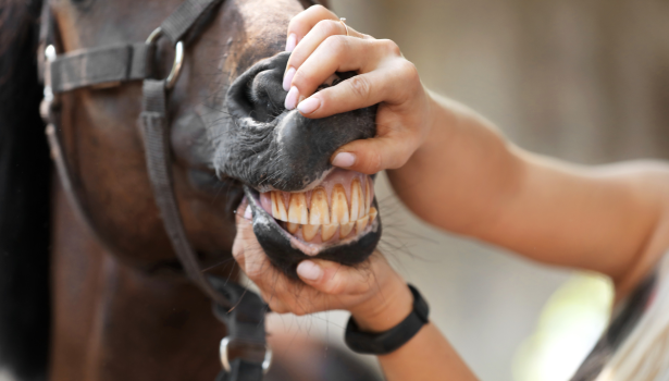
Wprowadzenie do stomatologii koni
Piśmiennictwo Loomans J.B.A., Stolk P.W.Th., van Weeren P.R., Vaarkamp H., Barneveld A.: A survey of the workload and clinical skills in current equine practices in The Netherlands. „Equine Veterinary Education”, 2007, 19: 162-168. Baker G.J., Easley J.: Equine dentistry. WB Saunders, 1999. Staszyk C., Wulff W., Jacob H.G., Gasse H.: The periodontal ligament of equine […]


Wskazówki na wypadek widocznego niepowodzenia terapii antybiotykowej. Kryteria skutecznej terapii oraz kluczowe pytania jako 5 kroków drzewa analitycznego
Czynniki powiązane z użyciem antybiotyku w terapii: Czy wybór antybiotyku opierał się na badaniach klinicznych i dodatkowych (diagnoza, antybiogram)? Sprawdź odpowiedź na pytanie 3. Farmakokinetyka/farmakodynamika wybranego antybiotyku? Koncentracja i czas działania antybiotyku w zakażonej tkance a efektywność w stosunku do czynnika bakteryjnego wywołującego chorobę (spektrum działania antybiotyku, wrażliwość z antybiogramu – odpowiedzi na pytanie 3.). […]


Czarno na białym – mastitis okiem praktyka – rozmowa z dr. n. wet. Sebastianem Smulskim
Rozmowa z dr. n. wet. Sebastianem Smulskim, pracownikiem Uniwersytetu Przyrodniczego w Poznaniu, specjalistą w dziedzinie profilaktyki i leczenia mastitis u krów, który w swoich badaniach zgłębia tematykę zapalenia gruczołu mlekowego u bydła, zarówno w aspekcie naukowym, jak i praktycznym. Większość zapaleń gruczołu mlekowego ma etiologię bakteryjną. Dlaczego, pomimo rozwoju mikrobiologii, medycyny weterynaryjnej i prowadzonych badań, […]


XVIII FORUM ZOOTECHNICZNO-WETERYNARYJNE: NOWE HORYZONTY W ROZRODZIE ZWIERZĄT
Na Uniwersytecie Przyrodniczym w Poznaniu w dniach 18-19 kwietnia br. odbyło się XVIII Forum Zootechniczno-Weterynaryjne pod hasłem „Rozród zwierząt w dobie selekcji genomowej”. To wydarzenie zgromadziło liczne grono lekarzy weterynarii oraz hodowców, by omówić najnowsze osiągnięcia w dziedzinie hodowli i rozrodu zwierząt. Organizacja Forum była wspólnym przedsięwzięciem Poznańskiego Koła Polskiego Towarzystwa Zootechnicznego, Wielkopolskiego Oddziału Polskiego […]


Wiosną zapisz się do bezpłatnego newslettera!
Bądź na bieżąco z nowościami z branży weterynaryjnej.