Chirurgia dróg oddechowych. Syndrom brachycefaliczny – cz. II
Respiratory surgery. Brachycephalic Airway Syndrome – Part II
Diagnosis of upper airway compromise can be made readily after taking a history, listening to the dog and conducting diagnostic imaging.
Clinical signs
Owners usually report heat, stress and exercise intolerance. Clinical signs include snoring, inspiratory dyspnea, cyanosis and in the most severe cases, syncopal episodes. Apneas can be observed during sleep (Farquharson and Smith, 1942).
Normally, during inspiration, the wings of the nostrils are actively dilated (Evans, 1993). If signs of dyspnea occur, the dogs stretch their necks upward to dilate the nasopharynx and, if necessary, to move the elongated soft palate below the epiglottis. When the elongated soft palate is held above the epiglottis, some dogs lie down on their sides, which releases the soft palate by allowing it to glide laterally to the epiglottis (Knecht, 1979; Singleton, 1962). In addition many owners describe signs of regurgitation, vomiting and dysphagia when their brachycephalic dog get excited or are in respiratory distress.
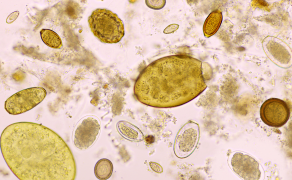
Diagnostic imaging
A proper evaluation of the airways should include neck and thoracic radiographs, computed tomography of the head and endoscopic examination of the upper airways, and if needed of the upper GI tract.
Radiologic examination
Thoracic radiographs are performed to document secondary heart or lung diseases. On occasion a sliding hiatal hernia can also be found on a lateral radiograph.
When CT scan is not available, a lateral radiograph of the neck can help to assess the soft palate thickness as defined by the soft tissue density present between the nasopharynx and oropharynx (Hendricks B., 1992).
Computer tomography
A computed tomography study of the skull allows an estimation of the level of obstruction and a detailed assessment of the nostrils, vestibule, nasal cavity, and naso-and oropharynx (Oechtering et al. 2007). The degree of vestibular stenosis, the ma...
którzy są subskrybentami naszego portalu.
i ciesz się dostępem do bazy merytorycznej wiedzy!

Zapisz się do bezpłatnego newslettera!
Bądź na bieżąco z nowościami z branży weterynaryjnej.